

IAP-26-046
Animal-free models to measure the impact of climate change on host-pathogen interactions in fish gills and skin
Infection is increasingly recognised as one of the dominant drivers through which climate change imposes pressure on fish biodiversity [1]. The mechanistic link is multi-faceted. At a physiological and cellular level, rising temperatures can increase pathogen metabolism and replication [2] and disrupt fish immune response [3]. From a spatial and ecological point of view, shifting geographic ranges favour encounters between pathogens and naïve hosts [4]. Behavioural adaptations, such as seeking cold refuges, increases fish concentration and contributes to the spread of disease [5]. Draught and low flow, particularly in freshwater environments, can increase pathogen concentration [6]. Such a diverse thematic landscape calls for novel multidisciplinary approaches that can guide and improve the efficacy of conservation efforts.
This PhD project focuses on understanding mechanistically how environmental changes impact fish-pathogen interactions at the level of cells and tissues. In particular, we will investigate two major interfaces that mediate contact with the environment: gills and skin. The student will be guided by an interdisciplinary supervisory team in leveraging new nanotechnological and imaging techniques to develop animal-free spatial models of these mucosal surfaces. This will allow us to visualize and quantify the cellular interactions between the fish immune system at the level of the epithelium and pathogens, with a particular focus on bacterial, fungal and protozoal species at different temperatures. The work will include live visualization and tracking of motile immune cells in the presence of pathogens. By achieving precise control over environmental conditions in the models, we will quantify the impact of climate change-associated changes in temperature, acidity, salinity, and UV exposure on these interactions. Our model system will be salmonids. Members of this versatile family are characterized by life histories that bridge sea and freshwater environments and that span a diverse range of developmental stages. This ecological and developmental breadth will allow us to establish principles that can be generalizable across environmental niches and host-pathogen interactions. We expect that our results will inform new targeted, directly implementable interventions for fish biodiversity conservation and provide new animal-free research approaches.
Objectives:
Aim1. Measure how environmental changes influence the behaviour of gill and skin pathogens.
Aim2. Develop an animal-free model of salmonid gills and skin and investigate how environmental changes affect tissue and cellular behaviour.
Aim3. Understand how environmental change affects the interactions between pathogens and salmonid immune cells.
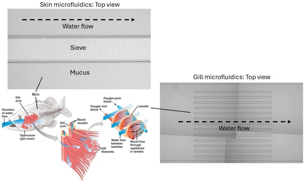
Click on an image to expand
Image Captions
Animal-free models of fish skin and gill models.
Methodology
We will use soft lithography and nanofabrication approaches available through the James Watt Nanofabrication Centre (JWNC) at the University of Glasgow to produce microfluidic and static imaging supports in which to interrogate host-pathogen interactions. The supports enable precise control over microenvironmental conditions, including flow rate, temperature, oxygenation, acidity, viscosity and nutritional profiles, allowing us to measure the impact of environmental changes associated to climate change. The environments will contain primary skin and gill epithelial cells, including skin keratinocytes. Co-culture with primary immune cells, including macrophages, B cells and T cells, will be explored and optimised. These will be challenged with bacteria (F. columnare, T. maritimum), oomycetes (Saprolegnia parasitica) or protozoal (Neoparamoeba perurans) pathogens both in isolation and in combination, allowing us to disentangle the impact of environmental change on each component of the host-pathogen interaction. Where possible, we will compare strains from culture collections to isolates maintained in the lab of ML. Salmonid cells will be provided by the lab of RRD. Quantification will be carried out through automated timelapse microscopy imaging and image analysis using dedicated equipment and code available in the lab of LM.
Project Timeline
Year 1
The first three months of the first year will focus on preparatory training. We do not expect this to cover the full spectrum of skills necessary for the project, but rather to provide a solid starting point on microbiology, keratinocytes and epithelial cell culturing, microfabrication, microscopy and image analysis. The student will receive specific training in the 3Rs framework (Replacement, Reduction and Refinement) and the relevant Home Office requirements, obtain a personal licence where required, and optimise cell isolation from humanely culled Atlantic salmon at HWU.
The training will then be integrated during the 3.5 years as required. For the remainder of the year, the student will initiate data collection on the behaviour of pathogens inside microfluidics that recapitulate certain aspects of the host environment (nutrients, spatial structure, fluid flow), but in the absence of the host (further referred to as “host-less” model).
Year 2
For the first three months of the second year the student will complete work on the “host-less” model, establishing a solid foundation that completes Aim1 and that will yield a first publication. Leveraging the skills acquired and complementing training with visits to the lab of RRD, the student will then produce an animal-free model of gill and skin. Similar to Aim1, they will study the impact of climate-relevant environmental changes on the model, using imaging and image analysis routines to quantify such changes. Focus will be particularly addressed to parameters such as metabolism, motility and growth.
Year 3
In the first three months of year three, the student will complete work on the gill and skin model for Aim2, which will yield a second publication. Leveraging the outputs generated until that time, the student will then move on to the third and final aim of studying the impact of environmental changes on host-pathogen interactions. The work carried out in Aim1 and 2 on the pathogen and host compartments in isolation will allow us to understand which behaviours emerge from interactions between them. Expected outputs include quantification of the dynamics of bacterial phagocytosis by skin keratinocytes, pathogen survival and killing, keratinocyte migration, epithelial damage, and pathogen colonisation success.
Results will be compared across climate change-relevant environmental conditions. This will allow us to determine mechanistically supported, environmental change-related risk factors that increase fish susceptibility to infection. Because we will investigate different pathogen families, our results will determine which infectious agents are most likely to pose risks to fish biodiversity and conservation.
Year 3.5
Work on Aim3 will be completed at the beginning of year 3, yielding a third publication. The 4 final months will be dedicated to thesis writing.
Training
& Skills
The student will be provided with interdisciplinary training in microbiology, fish tissue culture, imaging, soft lithography, microfluidics fabrication, and image analysis. The work will be carried out within the lab of LM in the School of Biodiversity, One Health and Veterinary Medicine, which will provide opportunities for training oral presentation skills through student-led seminars. Further scientific communication training will be afforded through attendance to conferences (International Society of Fish and Shellfish Immunology (ISFSI) Congress in 2028, MAST annual science meeting, The Fisheries Society of the British Isles Annual Symposium, Aquaculture Europe) and cross-laboratory presentation. Training in scientific writing and publishing will be supplied by the supervisory team ahead of publication. The student will have the chance to develop leadership and supervisory skills through the co-supervision of undergraduate and master’s projects.
References & further reading
1. 10.1016/j.envres.2024.120184.
2. 10.1111/raq.70112.
3. 10.1007/s10750-026-06120-3.
4. 10.1098/rstb.2013.0553.
5. 10.1111/jfd.14046.
6. 10.1111/1752-1688.12711
