

IAP-26-032
An investigation of interglobular dentine as a novel proxy for climate related childhood stress using XR-CT
Vitamin D is produced mainly through UV-B exposure, with a smaller dietary contribution from foods including fish and eggs, and is essential to immune and skeletal health. Deficiency records interactions between environment and society: irradiance, seasonality and latitude shape availability, while diet, mobility and child-rearing practices influence exposure. Prolonged childhood deficiency can cause rickets, impairing bone mineralisation and producing skeletal weakness and deformity; in adults it causes osteomalacia. Skeletal lesions may be subtle, remodel during growth or never become visible. Non-remodelling dental tissues therefore offer a more durable record of childhood deficiency. Interglobular dentine (IGD) forms when calcospherites fail to fuse during mineralisation, leaving hypomineralised zones along incremental growth lines. Associated with vitamin D deficiency, IGD can preserve the timing and recurrence of developmental disruption through the crown and root, potentially revealing episodes invisible in the skeleton. However, IGD’s diagnostic value remains unresolved. Research supports an association with rickets and low serum vitamin D, but no evidence-based threshold separates pathological from background levels, and its relationship with deficiency severity is unclear. Histology is destructive, captures a two-dimensional snapshot of a three-dimensional structure and commonly uses only qualitative scoring. Reproducible, non-destructive quantification would therefore be a major methodological advance.
This project will develop and validate a micro-CT and deep-learning workflow to detect and quantify IGD, asking: (1) can IGD be measured reproducibly from non-destructive three-dimensional images; (2) how does its distribution compare with skeletal and dental evidence of metabolic and developmental stress; and (3) did childhood stress vary among communities experiencing the AD 536–544 and other climatic disruptions?
The study will analyse teeth and skeletons from assemblages likely to have been affected by climatic disruptions. Combining dental microstructure, palaeopathology and biographical data, it will test how rapid environmental shock became embodied during childhood and whether vulnerability varied by age, sex and community. It links global environmental change with human health while delivering a transferable, non-destructive method for irreplaceable archaeological collections.
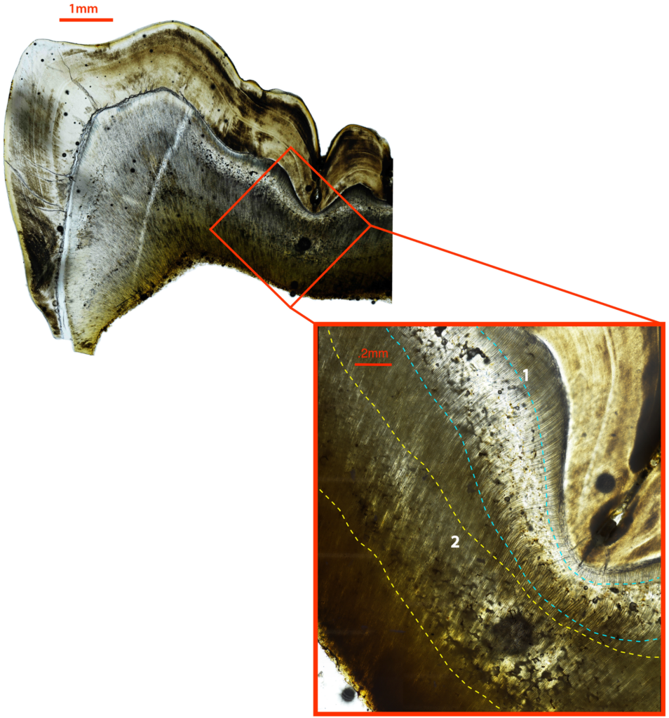
Click on an image to expand
Image Captions
Example of histological examination of IGD (Snoddy et al. 2024).
Methodology
The project will analyse 72 individuals from four securely dated sixth-century British assemblages (approximately 18 per site), selected for chronological and contextual variation. Sampling will prioritise individuals whose dental development can be related to AD 536–544 and who have associated skeletal data, including case studies previously analysed using histology (including by the supervisor, see Snoddy et al. 2024). Two permanent teeth spanning complementary developmental intervals will be selected per individual, with a third used selectively, producing 130–160 teeth. Pilot XR-CT scans will optimise resolution, contrast, scan time and radiation exposure across tooth types and preservation states before a standardised protocol is applied. Volumes will be reconstructed and processed in Dragonfly, with acquisition and processing parameters recorded for quality assurance and reproducibility.
A representative subset of 24–30 teeth will be independently annotated to train a supervised deep-learning model that segments IGD and quantifies its volume, density and distribution. Separate training, validation and held-out test sets, repeat annotation and comparison with expert observations and published histological criteria will assess accuracy and observer reliability. The validated workflow will then be applied to the remaining scans, with automated outputs quality checked. IGD will be compared with skeletal evidence of rickets or osteomalacia and indicators of childhood stress, including enamel hypoplasia, cribra orbitalia and porotic hyperostosis. Mixed-effects models will treat teeth as repeated measurements nested within individuals and test variation by site, developmental interval, tooth type, age and sex while accounting for preservation and sampling bias. Interpretation will require convergence among dental, skeletal and contextual evidence because IGD is not entirely specific to vitamin D deficiency. Age and sex will be estimated using multiple established indicators, and pathology recorded using standard guidance. A database will integrate context, chronology, preservation, demographic estimates, pathology and XR-CT measurements. Collection permissions and ethical requirements will be secured, and data, code and models documented and shared under FAIR principles.
Project Timeline
Year 1
Year 1: refine the research questions and sampling strategy; complete literature review; audit candidate assemblages and confirm chronology, secure ethical and institutional approvals; create the sampling and metadata database; undertake training in XR-CT and Python-supported image analysis; optimise scanning and quality-control procedures; present the protocol and prepare a methods or review paper.
Year 2
Year 2: undertake staged collection visits and sample transfer, recording provenance, condition, tooth selection and chain of custody; complete XR-CT acquisition for target 72 individuals and 130–160 teeth; complete macroscopic skeletal and dental recording; reconstruct and quality-check image volumes; create 24–30-tooth annotated dataset; develop, validate and document the segmentation workflow; undertake interim statistical analysis; present initial findings at UKAS conference.
Year 3
Year 3: apply validated workflow to full dataset and complete quality assurance; integrate tooth-level, individual, palaeopathological, chronological and contextual data; interpret assemblage-level and developmental patterns; prepare principal results papers alongside documentation of code, model information.
Year 3.5
Final six months: complete and submit thesis and publications; confirm that all physical material has been returned to collections.
Training
& Skills
· micro-CT acquisition, reconstruction, Three-dimensional image processing, expert annotation, deep-learning segmentation, model validation and reproducible computational workflows in Dragonfly and complementary tools including auditing appropriate Data Science modules.
· Quantitative analysis, including reliability testing, sensitivity analysis and integration of imaging, palaeopathological and contextual datasets.
Osteobiographical methods, including age and sex estimation, diagnosis of metabolic disease and childhood stress.
Research ethics, human-remains governance, FAIR data management, open research and responsible use of AI.
Project management, publication, conference presentation and communication with collection partners and public audiences.
References & further reading
Brickley, M. B., D’Ortenzio, L., Kahlon, B., Schattmann, A., Ribot, I., Raguin, E., & Bertrand, B. (2017). Ancient Vitamin D Deficiency: Long-Term Trends. Current Anthropology, 58(3), 420–427. https://doi.org/10.1086/691683
Colombo, A., D’Ortenzio, L., Bertrand, B., Coqueugniot, H., Knüsel, C. J., Kahlon, B., & Brickley, M. (2019). Micro-computed tomography of teeth as an alternative way to detect and analyse vitamin D deficiency. Journal of Archaeological Science: Reports, 23, 390–395. https://doi.org/10.1016/j.jasrep.2018.11.006
D’Ortenzio, L., Ribot, I., Raguin, E., Schattmann, A., Bertrand, B., Kahlon, B., & Brickley, M. (2016). The rachitic tooth: A histological examination. Journal of Archaeological Science, 74, 152–163. https://doi.org/10.1016/j.jas.2016.06.006
Helama, S., Arppe, L., Uusitalo, J., Holopainen, J., Mäkelä, H. M., Mäkinen, H., Mielikäinen, K., Nöjd, P., Sutinen, R., Taavitsainen, J.-P., Timonen, M., & Oinonen, M. (2018). Volcanic dust veils from sixth century tree-ring isotopes linked to reduced irradiance, primary production and human health. Scientific Reports, 8, 1339. https://doi.org/10.1038/s41598-018-19760-w
Snoddy, A. M. E., Shaw, H., Newman, S., Miszkiewicz, J. J., Stewart, N. A., Jakob, T., Buckley, H., Caffell, A., & Gowland, R. (2024). Vitamin D status in post-medieval Northern England: Insights from dental histology and enamel peptide analysis at Coach Lane, North Shields (AD 1711–1857). PLOS ONE, 19(1), e0296203. https://doi.org/10.1371/journal.pone.0296203
Veselka, B., Brickley, M. B., D’Ortenzio, L., Kahlon, B., Hoogland, M. L. P., & Waters-Rist, A. L. (2019). Micro-CT assessment of dental mineralization defects indicative of vitamin D deficiency in two 17th–19th century Dutch communities. American Journal of Physical Anthropology, 169(1), 122–131. https://doi.org/10.1002/ajpa.23819
